
Histotechnology Professionals Day is a celebration dedicated to the skilled individuals who turn the invisible world of tissues into vital information. While many people picture healthcare as doctors, nurses, and bustling hospital corridors, a quieter kind of expertise helps steer countless medical decisions: the careful preparation of tissue for microscopic review.
These professionals prepare microscopic tissue samples, revealing insights that can shape treatment decisions. A tiny biopsy or surgical specimen can hold the difference between “benign” and “needs urgent attention,” and that call depends on whether the tissue has been processed, sliced, and stained to show its story clearly.
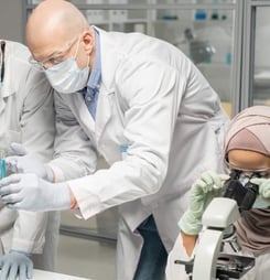
Through techniques like staining, sectioning, and embedding, histotechnologists reveal colors and details that help pathologists detect disease. Their work is largely unseen by the public, yet it plays a critical role in healthcare, guiding accurate diagnoses and ultimately impacting lives.
By honoring these specialists, Histotechnology Professionals Day brings attention to the essential work done behind laboratory doors. It shines a light on the commitment required to process samples correctly since even a minor error could alter a diagnosis.
It also highlights something easy to overlook: histotechnology is both science and craft. It requires knowledge of anatomy and chemistry, comfort with precision instruments, and the steady patience to repeat meticulous steps without cutting corners.
In a typical histology workflow, tissues are preserved so cells do not break down, processed so they can be supported by a firm medium, cut into sections thin enough for light to pass through, placed onto slides, and stained so structures become visible.
Depending on the case, that might involve routine stains such as hematoxylin and eosin (often called H&E), specialized stains that highlight particular tissue components, or immunohistochemistry methods that use antibodies to reveal specific proteins. Every step has a purpose, and each purpose supports the next set of eyes in the diagnostic chain.
How to Celebrate Histotechnology Professionals Day
Celebrating Histotechnology Professionals Day can be a fantastic way to honor these lab heroes who help diagnose illnesses with skill and precision.
The best celebrations are the ones that respect how labs function: safety matters, timing matters, and even well-meaning surprises should not disrupt a workflow built around patient care. With that in mind, these ideas keep things thoughtful, practical, and fun.
Show Appreciation with Goodies
Send a surprise treat to your local histology lab. A basket of snacks or a coffee delivery can brighten their day and keep them energized during long lab hours.
It’s a simple gesture, but it means a lot to those who work behind the scenes to support patient care. For a more lab-friendly approach, choose items that are easy to grab between steps, individually wrapped, and low-mess.
If the celebration is happening within a workplace, coordinating with lab leadership helps ensure treats arrive at a time that does not collide with critical tasks such as frozen section support, instrument maintenance, or slide deadlines.
Appreciation can also be practical. Restocking small comforts like hand lotion (frequent handwashing is real), quality pens for labeling, or a new box of lint-free wipes can feel oddly luxurious in a place where precision and cleanliness rule.
Even better is pairing the goodies with a note that acknowledges the work accurately: mention the attention to specimen identity, the skill of microtomy, or the expertise required to produce consistent staining.
Host a Mini Art Gallery
Set up a “tissue art” exhibit! Histotechnologists work with vibrant stains and slides daily.
Ask them to share some of their most stunning slides, then display these images in a mini-gallery at your workplace. This not only highlights their skill but also lets others see the artistry in histology.
Under the microscope, normal tissues can look like abstract landscapes, and special stains can produce bold color contrasts that feel almost painterly.
A mini gallery works best when it is paired with short, clear captions. The captions can explain what viewers are seeing in plain language: muscle fibers, glands, cartilage, or the architecture of skin layers.
If the lab is comfortable sharing educational images, the display can also describe what a stain is designed to highlight. This keeps the exhibit from being “pretty pictures only” and gently teaches how lab choices help reveal diagnostic clues.
For privacy and professionalism, the images should be de-identified and used with appropriate internal permissions. The point is to spotlight technique, not to tell anyone’s personal medical story.
Spotlight on Social Media
Post about the unsung heroes of healthcare on social media. Sharing posts, photos, and facts about histotechnology helps others learn about this fascinating field and the professionals who make it happen.
Use hashtags to connect with others celebrating the day and spreading awareness beyond the lab. Posts can highlight what histotechnologists actually do, since the job title is often unfamiliar outside of pathology circles.
A quick “day in the life” can be eye-opening: receiving and logging specimens, embedding tissue in paraffin blocks, cutting sections on a microtome, preparing special stains, and troubleshooting when a slide is too thick, too folded, or too pale.
If a workplace is participating, the most meaningful posts are often the ones that feature the people, with permission, and describe their expertise.
Even a short quote about what they enjoy, such as solving a staining problem or teaching a trainee how to face a block correctly, makes the profession feel real and human.
Encourage Educational Events
Invite a histotechnologist to speak at a local school or host a short webinar on histotechnology. These professionals can inspire students and curious minds with the science behind tissue analysis and pathology.
Educational outreach adds depth to the day and sparks interest in histotechnology careers. A well-structured talk can explain the big picture without overwhelming the audience: a physician collects a sample, a lab turns it into slides, a pathologist interprets those slides, and the results help guide care. From there, the speaker can introduce the core steps of tissue preparation and why each one matters.
Hands-on demonstrations can be simple and safe. A speaker might show a mock “embedding” with wax-like materials, explain how thin “sections” must be to allow light microscopy, or share microscope images of common tissues.
The goal is not to turn attendees into instant experts, but to show that this is a specialized, high-skill career that blends biology, chemistry, and problem-solving.
Organize a Thank-You Card Drive
Collect thank-you notes from coworkers, patients, or community members to show appreciation for the histotechnologists’ dedication.
Simple messages of gratitude can remind them that their work, though often unseen, makes a real impact on lives. The strongest messages are specific: thanking them for accuracy, consistency, and speed when it matters, or for the calm competence that keeps a lab running smoothly.
A card drive can also include “shout-outs” from pathologists, surgeons, and clinic staff. Many of those professionals rely on histology without ever seeing the fine details of how slides are made.
Encouraging them to name what they value, such as crisp margins on a surgical case or dependable immunohistochemistry results, reinforces that histotechnology is not an interchangeable step. It is an expertise.
For teams that want to go further, a card drive can turn into a recognition board in a break area, where colleagues post notes about problem-solving wins: fixing a difficult block, rescuing fragile tissue, improving a staining run, or training a new teammate. It becomes a living reminder that careful work is noticed.
History of Histotechnology Professionals Day
Histotechnology Professionals Day began in 2010, established by the National Society for Histotechnology (NSH). This day celebrates histotechnologists and their contributions to healthcare.
These specialists work in laboratories, processing tissue samples to help doctors diagnose diseases like cancer. Since this work often goes unnoticed, NSH wanted a day dedicated to raising awareness and showing appreciation for these lab professionals.
The timing of the day also reinforces its central message: histotechnology is essential to modern pathology and laboratory medicine, even if it is rarely visible to patients. In many healthcare journeys, the “answer” that shapes the next step is not a single lab number or imaging scan, but a pattern of cells on a glass slide. That slide exists because someone prepared it expertly.
Histotechnology itself has deep roots in medical science, growing alongside improvements in microscopes, staining chemistry, and laboratory methods. Over time, tissue preparation evolved from basic preservation and hand-cut slices into a sophisticated discipline.
Modern labs commonly use standardized processing equipment, precise microtomes, and validated staining methods so results are consistent across cases. Yet even with automation, histotechnology still depends on skilled judgment. Tissue is not uniform, and neither are clinical questions.
A histotechnologist may need to decide how to orient a specimen so the most important structures appear on the cutting surface.
They may adjust section thickness for delicate tissues, choose an appropriate stain to highlight a suspected organism or tissue component, or prepare slides for immunohistochemistry when a diagnosis requires specific markers. They also troubleshoot, because real specimens do not always cooperate.
A section may wrinkle, tear, or refuse to adhere to a slide. A stain may run light or show background. Fixation may be suboptimal, requiring extra care to produce interpretable results. These are not simply technical inconveniences. They can affect what the pathologist can confidently report.
Histotechnology Professionals Day also aligns with NSH’s broader interest in professional awareness and career development. The field depends on well-trained practitioners, and laboratories often need new talent with both technical ability and a steady approach to quality.
By calling attention to histotechnology as a career, the day supports the next generation of professionals who will keep diagnostic services running, train peers, and help labs adopt new methods.
The day’s history is therefore not only about recognition, but about visibility. When more people understand what histotechnologists do, it becomes easier to appreciate why labs emphasize careful specimen identification, consistent processes, and a culture of double-checking.
It also helps explain why histology can be time-sensitive: tissues must be preserved, processed, and stained with appropriate steps and timing to maintain morphology and, when relevant, antigen integrity for special studies.
Each year, the day allows histotechnologists, healthcare staff, and communities to come together, acknowledging the impact of this specialized work on patient outcomes.
It is a chance to celebrate the professionals who translate tissue into information, and to recognize that behind every clear slide is a person with practiced hands, sharp eyes, and the quiet pride of getting it right.